Introduction
Digital imaging has revolutionized the field of dentistry, providing dentists with advanced tools to enhance diagnosis and treatment. With the advent of digital imaging technology, traditional X-rays have been replaced by more accurate and efficient methods, allowing for better patient care and improved outcomes. In this article, we will explore the various digital imaging techniques used in dentistry and how they contribute to enhancing diagnosis and treatment.
Intraoral Cameras
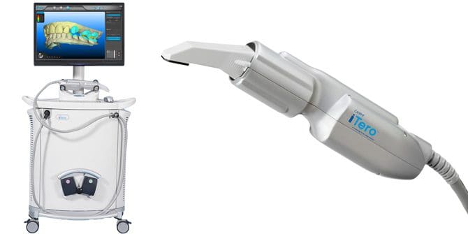
Intraoral cameras are small, handheld devices that capture high-resolution images of the inside of the mouth. These cameras allow dentists to examine the teeth, gums, and other oral structures in detail, helping them detect and diagnose various dental conditions. The images captured by intraoral cameras can be instantly displayed on a computer screen, enabling dentists to explain the findings to patients and develop personalized treatment plans.
Digital X-Rays

Digital X-rays have replaced traditional film-based X-rays in dentistry. These X-rays use electronic sensors to capture images of the teeth and supporting structures. Digital X-rays offer numerous advantages, including reduced radiation exposure, instant image availability, and the ability to enhance and manipulate images for better diagnosis. Dentists can zoom in, adjust contrast, and highlight specific areas of concern, aiding in the detection of cavities, bone loss, and other dental issues.
Cone Beam Computed Tomography (CBCT)
Cone Beam Computed Tomography, or CBCT, is a specialized imaging technique that provides three-dimensional images of the teeth, jawbone, and surrounding structures. CBCT scans are particularly useful for complex dental procedures such as dental implant placement, orthodontic treatment planning, and diagnosing temporomandibular joint disorders. The detailed images obtained through CBCT allow dentists to accurately assess bone density, identify nerve pathways, and plan treatments with precision.
Digital Smile Design (DSD)
Digital Smile Design is a cutting-edge technique that combines digital imaging and computer software to create a customized treatment plan for cosmetic dental procedures. Dentists can capture images of the patient’s face, teeth, and smile, and use specialized software to simulate the desired outcome.
Summary
Digital imaging in dentistry has transformed the way dental professionals approach diagnosis and treatment. By utilizing digital sensors and imaging software, dentists can capture detailed images of the teeth, gums, and surrounding structures. These images can be instantly viewed on a computer screen, enabling dentists to zoom in, enhance, and manipulate the images for a more comprehensive analysis.
One of the key advantages of digital imaging is its ability to reduce radiation exposure. Traditional X-rays required higher levels of radiation, whereas digital imaging systems use significantly less radiation, making it safer for both patients and dental professionals.
Furthermore, digital images can be easily stored, shared, and compared over time, allowing dentists to track changes in oral health and monitor the effectiveness of treatments. This not only improves patient care but also facilitates communication between dental specialists and other healthcare providers.
Overall, digital imaging has become an indispensable tool in modern dentistry. Its ability to provide high-quality images, reduce radiation exposure, and enhance diagnosis and treatment planning has revolutionized the field. A go to these guys s technology continues to advance, we can expect further improvements in digital imaging systems, leading to even better oral healthcare outcomes.
- Q: What is digital imaging in dentistry?
- A: Digital imaging in dentistry refers to the use of advanced technology to capture and store dental images electronically.
- Q: How does digital imaging enhance diagnosis and treatment?
- A: Digital imaging allows dentists to obtain high-quality images of the teeth, gums, and other oral structures, enabling more accurate diagnosis and treatment planning.
- Q: What are the benefits of digital imaging in dentistry?
- A: Some benefits include reduced radiation exposure, immediate image preview, easy storage and retrieval of images, and the ability to enhance and manipulate images for better analysis.
- Q: What types of digital imaging techniques are used in dentistry?
- A: Common digital imaging techniques in dentistry include intraoral cameras, digital X-rays, cone beam computed tomography (CBCT), and 3D imaging.
- Q: Is digital imaging safe?
- A: Yes, digital imaging in dentistry is considered safe. It uses lower radiation doses compared to traditional X-rays and follows strict safety protocols to minimize any potential risks.
- Q: How are digital images stored and shared?
- A: Digital images are typically stored in secure electronic databases or cloud-based systems. They can be easily shared with other dental professionals or specialists for collaborative treatment planning.
- Q: Can digital imaging help with cosmetic dentistry procedures?
- A: Absolutely. Digital imaging allows dentists to create virtual smile makeovers, simulate the results of cosmetic procedures, and help patients visualize the potential outcomes before treatment.
- Q: Are there any limitations to digital imaging in dentistry?
- A: While digital imaging offers numerous advantages, it may not be suitable for every dental condition. In some cases, traditional diagnostic methods or additional tests may still be necessary for a comprehensive evaluation.

Welcome to my website! My name is Charles Boyland, and I am a dedicated professional Orthodontic Consultant with a passion for promoting oral health and providing innovative dental solutions. With years of experience in the field, I am committed to helping individuals of all ages achieve a healthy and confident smile.

